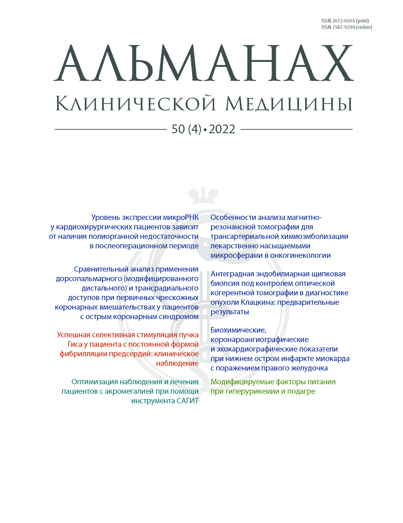
Vol 50, No 4 (2022)
- Year: 2022
- Published: 29.11.2022
- Articles: 8
- URL: https://almclinmed.ru/jour/issue/view/72
Full Issue
ARTICLES
The level of microRNA expression in cardiac surgery patients depends on postoperative multiorgan failure
Abstract
Aim: To assess the level of microRNA expression in the serum of patients who had undergone cardiac surgery depending on the postoperative complications (presence or absence of multiorgan failure, MOF).
Materials and methods: The study group included 87 patients who had undergone heart surgery with cardiopulmonary bypass. The patients without postoperative complications comprised group 1 (n = 51), whereas those with postoperative MOF were in group 2 (n = 36). In all patients, blood samples were collected at two time points: before surgery and at 36 to 48 hours after surgery. The following miRNAs were chosen for the study: hsa-miR-486-5p (478128_miR), hsa-miR-191-5p (477952_miR), hsa-miR-192-5p (478262_miR), hsa-miR-146a-5p (478399_miR), hsa-miR-26a-5p (477995_miR), hsa-miR-30d-5p (478606_miR), hsa-miR-23a-3p (478532_miR), and hsa-miR-320a-5p (481049_miR). Polymerase chain reaction results were normalized to hsa-miR-16-5p (4427975).
Results: Up-regulating miRNAs. Compared to baseline, there was a significant postoperative increase in miR-486-5p microRNA expression (group 1, 41.83 [19.86; 74.6] vs 940 [434.7; 1212.0]; group 2, 72.55 [21.37; 100.2] vs 492.4 [201.2; 998.0]; both p < 0.001). An increase in the of microRNA miR-192-5p expression in the postoperative period was found both in the no-MOF group (from 0.39 [0.16; 1.07] at baseline to 5.96 [3.74; 10.35] after surgery, p = 0.002), and in the MOF group (from 1.74 [0.45; 3.35] at baseline to 17.16 [4.70; 24.96] after surgery, p = 0.003), with a statistically higher level of expression in group 2 (p = 0.028). Similar changes over time were observed for miR-30d-5p expression: group 1, 1.61 [0.47; 4.36] at baseline vs 5.03 [2.93; 6.56] after surgery (p = 0.002), group 2, 0.89 [0.32; 4.27] at baseline and 6.63 [3.92; 12.82] after surgery, respectively (p = 0.0045).
Down-regulating miRNAs. The miR-191-5 and miR-146a-5p families demonstrated a significant increase in group 1 after surgery (3.85 [1.64; 5.6] vs 7.7 [5.48; 9.68], p = 0.021; and 18.1 [6.52; 19.9] vs 37.27 [29.13; 47.07], p = 0.016, respectively) and a significant postoperative decrease in group 2 (3.67 [2.60; 7.61] vs 1.66 [0.52; 2.36], p = 0.023; and 14.75 [12.79; 21.77] vs 5.96 [2.8; 8.2], p = 0.034, respectively), with between-group difference in the postoperative expression levels being also significant. As regards to miR-26a-5p и miR-23a-3p families, there was a similar trend: the group with uncomplicated postoperative course was had virtually no changes over time in their expression (the increase was non-significant), whereas the MOF group had lower postoperative values for this microRNA family. The between-group differences after surgery were significant for miR-26a-5p (group 1, 6.79 [3.38; 8.46], group 2, 0.26 [0.18; 1.9], p = 0.037) and for miR-23a-3p (14.14 [11.92; 26.63] and 2.0 [1.02; 4.18], respectively, p < 0.001).
Conclusion: When comparing microRNA expression before surgery, we did not find any significant differences between the patients groups without and with MOF. Assessment of microRNA expression in the no-MOF group after surgery showed an increase in the expression of microRNAs responsible both for up regulation (miR-486-5p, miR-192-5p, miR-30d-5p) and for down regulation (miR-191-5p, miR-146a-5p). In the group with a complicated postoperative course and with MOF, changes over time in the up-regulating microRNAs were characterized by increased expression (miR-486-5p, miR-192-5p, miR-30d-5p), whereas the down-regulating microRNAs (miR-191-5p, miR-146a-5p) demonstrated significantly decreased expression, which was different both that in the no-MOF group and from the baseline values.
 217-225


Specific characteristics of the magnetic resonance imaging for transarterial chemoembolization with drug-saturated microspheres in oncogynecology
Abstract
Background: Magnetic resonance imaging (MRI) is used for the staging and assessment of treatment results of female pelvic tumors. The inclusion of transarterial chemoembolization (TACE) with drug-saturated microspheres into the treatment regimen puts a question to the radiologist: what TACE characteristics should be taken into account for the correct interpretation of the treatment results?
Aim: To determine the main MRI parameters that characterize the results of TACE in the treatment of women with primary and recurrent pelvic tumors.
Materials and methods: We performed a retrospective observational study of 80 patients with primary tumors (group 1) and 20 patients with recurrent tumors (group 2) of the small pelvis, complicated by tumor bleeding, who underwent 121 TACE procedures from 01.09.2015 to 01.12.2021 and were followed up to May 31, 2022. The study inclusion criteria were as follows: compliance with the approved protocol and time points for pelvic MRI. TACE results were evaluated according to RECIST 1.1.
Results: In 100% of the cases in the groups 1 and 2, bleeding was controlled within 24 hours. In group 1, partial response was achieved in 48% (n = 38), complete response in 15% (n = 12), stabilization in 37% (n = 30), without any progression in all patients. In group 2, partial response was achieved in 27% (n = 5), complete response in 11% (n = 2), stabilization in 62% (n = 13), without any progression, as well. When comparing the mass volumes, recurrent tumors were significantly more responsive to TACE. The type of tumor growth was infiltrative (n = 25), expansive (n = 55), and mixed (n = 20). No significant differences in volume changes depending on the type of tumor growth were found. Eight women had undergone non-targeted ovarian embolization related to the type of blood supply. There were no cases of non-targeted embolization of the abdominal organs and the bladder, even with existing abnormal collateral vasculature.
Conclusion: According to this data, the results of TACE for primary and recurrent pelvic tumors are characterized with the following MRI parameters: 1) hemostatic and cytostatic effects of TACE are manifested independently of each other; 2) tumor volume reflects changes after TACE to a greater extent than changes in linear dimensions; 3) there are cases of non-targeted ovarian embolization.
 226-236


Antegrade endobiliary forceps biopsy under the optical coherence tomography control in the diagnosis of Klatskin tumor: preliminary results
Abstract
Background: Transcutaneous transhepatic endobiliary forceps biopsy is an accepted method for verification of extrahepatic cholangiocarcinoma, but its sensitivity ranges from zero to 94%. In the recent years, optical coherence tomography (OCT) has been actively used to diagnose malignancies.
Aim: To assess diagnostic accuracy of the OCT-assisted intraductal forceps biopsy in patients with Klatskin tumor.
Materials and methods: From 2013 to 2021, 161 patients with preliminary diagnosis of Klatskin tumor were seen in Russian Scientific Center of Radiology and Surgical Technologies named after Academician A.M. Granov. The retrospective study included 48 patients and 51 procedures of the forceps biopsy. In 14 (29%) patients of the main study group, the biopsy procedure was performed with OCT assistance, whereas the control group (34 patients, 71%) had their biopsies without the OCT.
Results: All procedures were technically successful. In the main and in the control study groups, sensitivity was 92.3% versus 73.3% (p = 0.32) and specificity 100% versus 85.7% (p = 0.88), respectively. Malignant neoplasm of the biliary tract was found in 13 cases versus 23 in the control group, with the degree of the tumor differentiation being determined in 64.3% (n = 9), versus 48.7% (n = 18) (p = 0.89), respectively. There were no adverse events associated with OCT and biopsy sampling in the main study group. In the control group, 4/37 procedures (10.8%) were associated with hemobilia, which was successfully treated conservatively within 24 hours without any prolongation of the hospital stay.
Conclusion: Our preliminary results indicate that antegrade endobiliary forceps biopsy is a safe and informative technique. The OCT navigation increases the sensitivity and specificity of the diagnosis. This allows for a personalized choice of chemotherapy. OCT is a promising technique for differential diagnosis of Klatskin tumor from benign biliary strictures. Further large-scale studies are required to introduce it into everyday practice.
 237-244


Comparative analysis of the dorsopalmar (modified distal) and transradial access in primary percutaneous coronary interventions in patients with acute coronary syndrome
Abstract
Background: Primary percutaneous coronary interventions (PCI) in acute coronary syndrome (ACS) with transradial access (TRA) are associated with the risk of local complications, such as occlusion of the radial artery (ORA), hematomas, pseudoaneurysms, and arteriovenous fistulas.
Aim: To perform comparative assessment of clinical efficacy and safety of the TRA and dorsopalmar (modified distal) radial access (DpRA) for primary percutaneous coronary intervention in in-patients with ACS.
Materials and methods: This was a randomized, dynamic, single-center, prospective study in two parallel groups. The patients were randomized in a 1:1 ratio into two groups with different types of the radiation access: TRA (n = 100) or DpRA (n = 100). TRA was made at the distal third of the forearm and DpRA on the dorsal palm surface. After the access zone was evaluated by angiography, the pressure bandage was placed on the zone for 6 hours for hemostasis. The comfort of hemostasis was assessed by the Gaston-Johansson 10-point verbal-descriptive pain rating scale. On the 5–7th day after PCI, all patients were examined with palpation and ultrasound assessment of the access artery.
Results: The number of attempts, average duration of the radial artery puncture, duration of the fluoroscopy procedure, and the conversion rate did not depend on the access type. The scoring of the subjective hemostasis comfort showed a significant advantage of DpRA over TRA (6.4 [4; 10] in the TRA group vs 1.7 [0; 6] in the DpRA group, p < 0.001). The rate of EASY III hematomas was 15 (15%) in the TRA group vs 3 (3%) in the DpRA group (p = 0.004). There were no EASY IV–V hematomas, occlusion of the radial artery of the forearm, pseudoaneurysms and arteriovenous fistulas in the DpRA group. The diameter of the forearm radial artery was significantly larger than the diameter on the dorsal palm surface in the patients of both groups, regardless of the type of access chosen (2.75 ± 0.32 mm and 2.38 ± 0.36 mm in the TRA group, p < 0.001; 2.84 ± 0.38 mm and 2.45 ± 0.36 mm in the DpRA group, p < 0.001). In the patients with access conversion in both groups, the diameter of the radial artery at both levels was less than the average one.
Conclusion: DpRA for PCI in ACS patients is a safe alternative to conventional radiation access. Ultrasound examination of the radial artery diameter in its distal and forearm parts before PCI could reduce the conversion rate.
 245-254


Biochemical, coronary angiographic and echocardiographic parameters in inferior acute myocardial infarction with right ventricle injury
Abstract
Background: The involvement of the right ventricular (RV) myocardium in inferior acute myocardial infarction (AMI) increases the risk of complication and death rates, which makes it important to timely identify this type of myocardial infarction.
Aim: To assess the value of functional, biochemical, coronary angiographic and ultrasound parameters in the patients in their productive age with inferior AMI, in order to identify the RV injury before and after percutaneous coronary intervention (PCI).
Materials and methods: This cohort prospective study included 141 patients with inferior AMI and ST elevation (26 women aged up to 60 years and 115 men aged up to 65 years), admitted to the emergency of the Medical Unit of Kazan (Volga region) Federal University from 2019 to 2021. The patients past history, clinical, biochemical and ultrasound data were obtained on admission and at discharge from the hospital. The two-dimensional speckle tracking echocardiography was performed at days 5 to 7 after PCI. The results are given as median values and 25% and 75% quartiles (Ме [Q1; Q3]).
Results: According to electrocardiographic signs, 41.8% (n = 59) patients with inferior AMI comprised the group with the RV injury. There were no differences in the myocardial injury biomarker levels between the groups on admission (р = 0.31 and p = 0.786, respectively). The coronary angiography showed that the index artery was the right coronary artery in 100% (n = 59) cases with the RV injury and in 67.1% (n = 55, р < 0.001) of the cases without the RV injury. Proximal involvement was 2.7 more common in biventricular infarction, than in the isolated inferior one (р = 0.013). During PCI, the RV involvement significantly increased the risk of complications (in 28 (47.5%) and 18 (22.0%) of the cases, respectively, р < 0.001), among them being the need in a temporary pacemaker placement (8 (13.6%) and 2 (2.4%) patients, р = 0.027). Echocardiography showed worse parameters of global and local contractility of both ventricles in the group with the RV involvement in the inferior AMI. The left ventricular (LV) ejection fraction decreased from 55% [51; 57] to 52% [47; 56] (р = 0.005); global RV deformity from -15.2% [-18.5; -13.4] to -12.3% [-15.6; -10.6] (р < 0.001); total number of segments with local contractility abnormalities increased from 2 [1; 3] to 5 [3; 6] (р < 0.001).
Conclusion: The study has confirmed that the involvement of RV into inferior LV AMI in the patients of productive age should be verified by abnormalities of electrocardiographic, biochemical, coronary angiographic and ultrasound parameters. To document the RV injury before PCI, ST elevation in additional right chest leads (V3R–V4R) was most informative, whereas after PCI, it was the finding of abnormal local contractility of basal and medial inferior RV segments by two-dimensional echocardiography and decreased longitudinal RV deformation by speckle tracking.
 255-263


REVIEW ARTICLE
Modifiable nutritional factors in hyperuricemia and gout
Abstract
Uric acid is an independent risk factor for socially important diseases, such as chronic heart failure and chronic kidney disease. The review summarizes current data on the impact of nutrition as a presumably modifiable factor into the development of hyperuricemia and gout. The authors describe biochemical mechanisms underlying hyperuricemia and present data on the influence of excessive intake of fructose, purines, and various alcohol-containing drinks on the risk of gout. Favorable impact of vitamin C supplements, coffee, cherry juice, some chemical elements (magnesium, zinc, and copper) on uric acid levels has been shown, as well as the impact of intake of low fat dairy products, polyphenols, food fibers, and omega-3 polyunsaturated fatty acids on the incidence of hyperuricemia. The information presented in the review can be used in clinical practice for preparation of an individualized nutritional plan aimed at normalization of uric acid levels. This would minimize the use of agents affecting the synthesis of uric acid and slow down the progression of chronic heart failure and chronic kidney disease.
 264-273


LECTURE
Optimization of the follow-up and treatment of patients with acromegaly with the SAGIT® tool
Abstract
The condition of a patient with acromegaly is significantly influenced not only by growth hormone (GH) and insulin-like growth factor type 1 (IGF-1) levels, but also by specific characteristics of somatotropinoma, severity of the main symptoms of acromegaly, and concomitant disorders. After successful achievement of biochemical control (GH and IGF-1 goals) the expected life longevity in patients with acromegaly could be comparable with that in the general population. It is important to avoid therapeutic inertia, to achieve biochemical control within shortest time period and to carefully tailor the treatment of concomitant disorders.
The cumulative experience of patient management has shown that therapeutic decisions should be based not only on measurement of GH and IGF-1 levels, but also on a unified multifactorial assessment of the patient's status. For this purpose, the SAGIT® tool has been elaborated, which ensures a multifaceted standardized assessment of patients with acromegaly, taking into account clinical symptoms, hormonal parameters, tumor size and comorbidities. This paper is the first description of the SAGIT® tool in Russian. The use of this tool allows for standardization of the patients assessment and for comparison of the status in patients both within one center and in different centers. The key unified information obtained with SAGIT® could be the basis for making prompt decisions on the need in treatment optimization for a patient with acromegaly, including his/hers referral to an expert center for the choice of further treatment strategy.
The SAGIT® tool has been recommended for implementation into clinical practice by international professional associations. It is advisable to perform studies with participation of the Russian patients with acromegaly to assess the possibility of SAGIT® implementation into clinical practice in Russia.
 274-280


CLINICAL CASES
The successful selective His bundle pacing to the patient with permanent atrial fibrillation
Abstract
The choice of an optimal, most safe and physiological place in the heart for the electrode implantation is an issue to be solved by a specialist in surgery of heart arrhythmias. According to the literature, the techniques to stimulate both cardiac apex and other alternative areas are imperfect. At present, stimulation of the heart conduction system, namely, the His bundle is considered a promising area in arrhythmology. This type of stimulation is a physiological one, while it involves the His-Purkinje system. We present a successful clinical case of the electrode implantation with a two-chamber pacemaker to the heart conduction system in a 67-year-old patient with permanent atrial fibrillation. The surgical intervention made it possible to reduce the QRS length from 180 to 110 ms. This clinical case shows that the heart conduction system pacing allows for implementation of the principles of physiological stimulation in patients with impaired atrioventricular conduction. The technique with a two-chamber pacemaker is feasible in clinical practice.
 281-285


