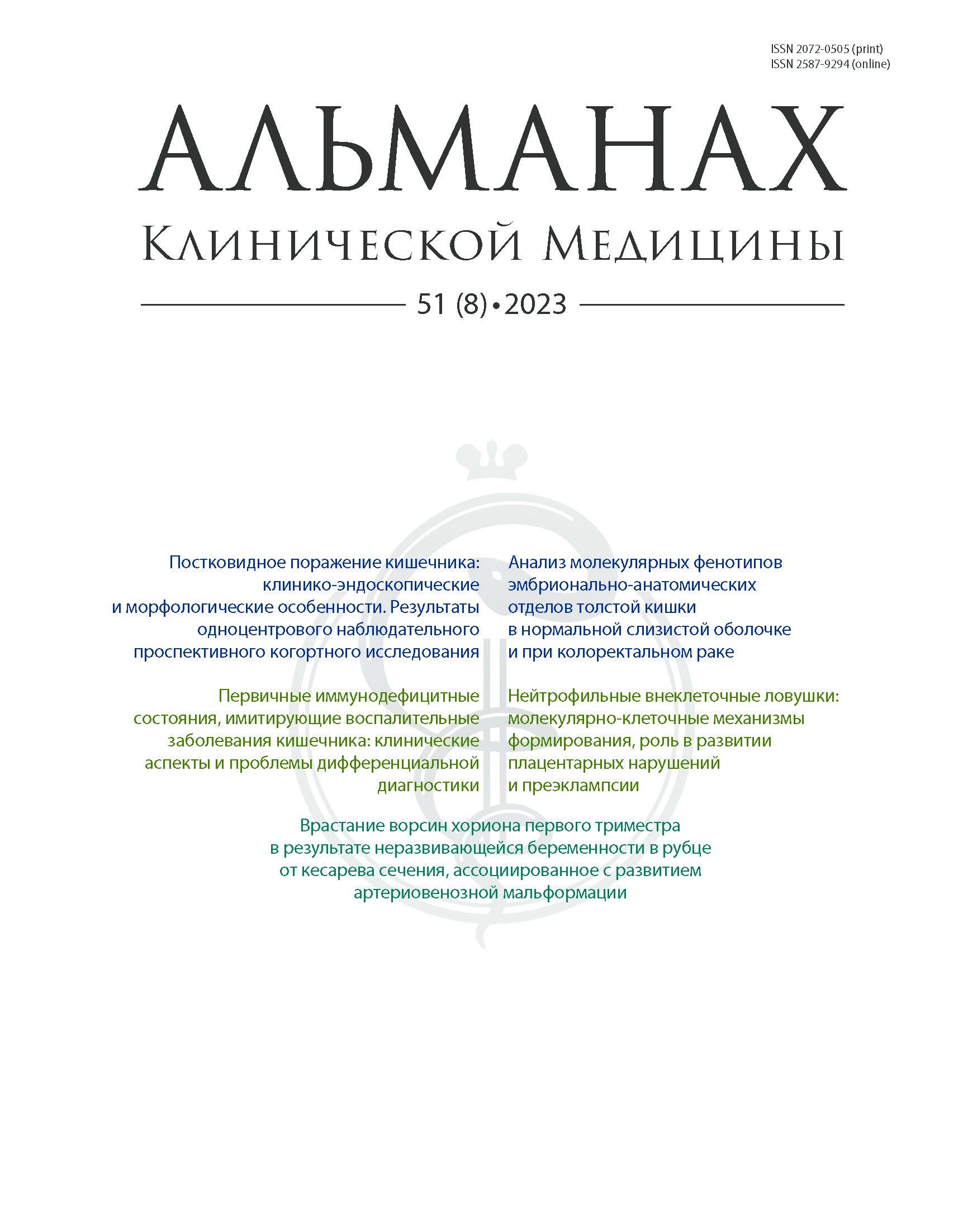
Vol 51, No 8 (2023)
- Year: 2023
- Published: 11.12.2023
- Articles: 6
- URL: https://almclinmed.ru/jour/issue/view/85
Full Issue
ARTICLES
The post-COVID-19 intestinal damages: clinical, endoscopic and morphological features. The results of a single-center prospective observational cohort study
Abstract
Background: Clinical signs of gastrointestinal disorders can manifest both from the first days of COVID-19 and after recovery, and may last up to 6 months or more. Most studies examined the gastrointestinal changes in acute coronavirus infection, whereas intestinal abnormalities in the early and late post-COVID period and their causes have not been sufficiently studied.
Aim: To determine the frequency and types of clinical, endoscopic and morphological abnormalities in patients with post-COVID-19 intestinal lesions.
Materials and methods: This was a prospective, observational, open-label, cohort, non-controlled study in 72 patients with intestinal symptoms after the coronavirus infection (female 48, mean age 54.6 (95% confidence interval 51.08–58.12) years), who were admitted to the Department of Gastroenterology of general hospital during the first and second waves of COVID-19 from June 2020 to September 2021. The assessment included routine anamnestic, clinical, laboratory, endoscopic, and morphological methods. When indicated, visualization methods (ultrasound, computed tomography, magnetic resonance imaging) were performed. The treatment was symptom-oriented and aimed at inflammation, anemia and protein and electrolyte abnormalities. Outcomes were assessed by the time of discharge from the hospital and thereafter by telephone interviewing of the patients for 8 weeks.
Results: In all patients, the main symptom was diarrhea, which started right just after SARS-CoV-2 infection with negative PCR test or 2–4 weeks later. The average stool frequency was 6.8 (5.61–7.99) times daily. In 19/72 patients (26.4%), there were blood and mucus in stools. 2.8% of the patients developed massive intestinal bleeding. Fever was present in 40.3% of the patients, decreased hemoglobin levels in 44.4%, and hypoalbuminemia in 16.7%. Signs of systemic inflammation (increased erythrocyte sedimentation rate, C-reactive protein, fibrinogen, thrombocytosis, leukocytosis) were found with various frequencies in a half of the patients. Clostridioides difficile A and B toxins were identified in 38.9% of the cases and increased fecal calprotectin in 22.2%.
Ileocolonoscopy was performed in 67 patients. The colonic mucosa in 21 (31.3%) patients either was not different from the normal, or showed minimal inflammatory changes such as absence of vascular pattern, hyperemia, mild friability even in patients with severe diarrhea, fever and laboratory abnormalities. Pseudomembranous colitis was diagnosed in 12 (17.9%) patients, and focal hemorrhagic colitis in 11 (16.4%) patients. In 2 (3%) cases, moderate to severe ulcerative colitis was newly diagnosed after the SARS-CoV-2 infection. Single or multiple erosions and ulcers of various sizes against the unchanged surrounding mucosa were found in 19 (28.4%) patients. In 2 (3%) cases with profuse intestinal bleeding, the endoscopy showed diffuse spontaneous bleedings from colonic mucosa, with no local source of bleeding found.
Biopsy samples from colonic mucosa were taken from 47 patients. Morphological abnormalities of only moderate or dense lymphoplasmocytic infiltration were detected in 29 (67.7%) patients, erosions in 8 (17%). In 2 (4.3%) cases, highly dense lymphoid infiltration with neutrophils, eosinophils and crypt abscesses was found, as typical for ulcerative colitis. In 2 (4.3%) patients, the lymphoid infiltration was associated with small venular and arteriolar clots, and in 5 (10.6%) patients there were no significant morphological abnormalities, except mild lymphoid infiltration.
During the hospitalization period, 56.9% of the patients showed a decrease in stool frequency, improvement of clinical and laboratory signs of systemic inflammation and metabolic abnormalities, and partial restoration of their body weight. One patient with ulcerative colitis received adalimumab and achieved stable clinical and endoscopic remission. Complete recovery was noted in 27.8% of the cases within 4–8 weeks, among them in 1 patient who had had profuse intestinal bleedings. In 3 cases (4.2%), an emergency colectomy was performed due to severe pseudomembranous colitis. Two (2.8%) deaths were registered: one patient with newly diagnosed post-COVID severe ulcerative colitis died of pneumonia, and another patient died of severe multiorgan failure. In 6 (8.3%) cases, a moderate long-term (above 8 weeks) diarrhea with metabolic disorders, weight loss, but without systemic inflammation was noted.
Conclusion: In patients with post-COVID intestinal lesions, the clinical symptoms are almost the identical and manifest with diarrhea of varying severity, systemic inflammation and metabolic disorders, whereas the endoscopic and morphological lesions are quite diverse. The severity of clinical symptoms often does not correspond to poorly expressed endoscopic and morphological signs. The variability and inconsistency of the clinical, endoscopic and morphological changes have not yet been systematized and do not allow for a clearly formulated diagnosis (except cases of pseudomembranous and ulcerative colitis). This makes it impossible to provide pathophysiologically-based treatments.
 427-440


Analysis of molecular phenotypes in normal mucosa and colorectal cancer in embryonic anatomical parts of the colon
Abstract
Background: Differences in the embryonic development of the colonic mucosa determine the physiological embryonic-anatomical asymmetry of its structure and can manifest themselves via different molecular phenotypes (expression profiles) of the colon segments. These molecular characteristics are hypothesized to determine differences in the carcinogenesis mechanisms and influence the prognosis of right- or left-sided colorectal cancer (CRC). Studies of the tumors molecular phenotypes depending on their localization may be of interest for assessment of the prognosis and choice of treatment for CRC.
Aim: To perform comparative analysis of molecular phenotypes of the normal colonic mucosa and adenocarcinoma CRC tissues depending on the natural embryonic anatomic asymmetry of the colon.
Materials and methods: We performed a retrospective study of molecular phenotypes (mRNA expression of 61 genes) from different embryonic-anatomical parts of healthy colon and CRC. The normal group included 254 samples of mucosa from three different parts of the colon from 74 healthy donors who had no cancer and no organic abnormalities of the colon, including 90 samples from the right colon, 116 from the left colon, and 48 from the rectum. The CRC group consisted of 154 samples of localized stage T1–4N0–2M0 adenocarcinoma from 154 patients who had not received neoadjuvant radio- and chemotherapy, including 40 samples from the right colon, 54 from the left colon, and 60 from the rectum. The relative mRNA abundance of 61 genes was assessed by reverse-transcriptase polymerase chain reaction. In both groups, the resulting expression phenotypes were compared between the anatomical parts of the colon. Statistical management of the data included the discriminant analysis with stepwise inclusion of variables.
Results: Based on the assessment of the mRNA level of the studied genes, a discriminant model was built that allows for classification of the normal group samples according to their anatomic origin in the colon with an accuracy of 95.8%. The most significant (p < 0.05) for classification are the following 19 genes: CCND1, SCUBE2, TERT, BAG1, NDRG, IL1b, IL2Ra, IL7, ESR1, TGFb, IGF1, MMP9, MMP11, PAPPA, CD45, CD69, TLR2, TLR4, LIFR. The discriminant model built for the CRC group included 27 genes and made it possible to differentiate samples from three parts of colon with an accuracy of 75.2%. A statistically significant (p < 0.05) contribution to the samples differentiation by the discriminant model was made by the COX-2, BIRC5, LIFR, TPA, IL1b, MMP11, MMP7, and P16INK4A genes. When combining samples from the two groups into one model in accordance with their embryonic-anatomical origin, there was a clear separation of tumor tissue samples and healthy colonic mucosa in the discriminant function space.
Conclusion: The analysis of CRC gene expression profiles using the discriminant model showed that genetic changes in the colonic mucosa in CRC flatten the molecular phenotypic boundaries of the embryonic-anatomical parts. These changes are specific to CRC, forming a particular “pathological” molecular phenotype.
 441-455


REVIEW ARTICLE
Primary immunodeficiency disorders imitating inflammatory bowel diseases: clinical aspects and problems of the differential diagnosis
Abstract
From the beginning of 2000s, there has been a significant increase in the incidence of inflammatory bowel diseases (IBD) and primary immunodeficiency disorders (PIDs) in adults and children in many countries around the world. The aim of the review is to summarize the state-of-the-art on diverse clinical types of PIDs with gastrointestinal manifestations and their differential diagnostic algorithms.
Atypical PIDs with “blunted” clinical manifestations are challenging for the timely diagnosis. Some types of PIDs with gastrointestinal involvement are also difficult to differentiate with classical IBDs. Molecular genetic studies have allowed for selection of a specific group of monogenic IBD-like diseases, represented mainly by PIDs. The authors discuss current classification of PIDs and their main clinical types imitating IBD, with important clinical and laboratory aspects. High level of information and awareness of practicing specialists working with IBD patients would be helpful in the selection of a patient cohort with possible PIDs and in the performance of extended laboratory assessment or referring for genetic tests. Timely diagnosis of PIDs would ensure quick administration of target therapy or hematopoietic stem cell transplantation, which in most cases would allow for the achievement of the disease remission, improvement of quality and duration of life.
 456-468


Neutrophil extracellular traps: molecular and cellular mechanisms of formation, role in the development of placental disorders and preeclampsia
Abstract
The review summarizes current understanding of neutrophil extracellular traps (NETs) and their role in the development of inflammation and thrombus formation during physiological and complicated pregnancy. The main initiation factors and molecular and cellular reactions leading to the generation of NETs are described. During gestation, various pregnancy-associated triggers (cytokines, hormones, colony-stimulating factors, etc.) contribute to increased activity of innate immune factors associated with the processes of neutrophil migration into gestational tissues, adhesion, degranulation, phagocytosis and release of extracellular neutrophil traps. It has been established that the uncontrolled aberrant generation of NETs, as well as their products, including reactive oxygen species, can exert a cytotoxic effect on maternal cells and tissues, adverse fetal effects and contribute to placental damage, resulting in such pregnancy complications as placental disorders, immunothrombosis and preeclampsia. The emergence of new data on the morphological and functional characteristics of the cellular component of innate immunity necessitates their advanced research with consideration of the functional potential and conditions for NETs formation, clarification and determination of their pathophysiological significance in normal and complicated pregnancy. It seems promising to study the possibility of assessment of the DNA traps levels for early diagnosis and prognosis of gestational complications, as well as for the development of new treatment strategies including targeted therapy.
 469-477


CLINICAL CASES
Ingrown chorionic villi of the first trimester as a result of a non-developing pregnancy in the post-cesarean scar, associated with the development of arteriovenous malformation: а clinical case
Abstract
Background: Identification of residual chorionic tissue and ingrowing chorionic villi after uterine cavity curettage due to non-developing pregnancy, spontaneous abortions, and medical abortions has been a poorly studied problem. The most challenging is the differential diagnosis of this condition when the chorion grows into the scar from a caesarean section and is associated with arteriovenous malformations of the uterine wall. Nowadays, ultrasound has been recognized as the primary diagnostic method; however, the absence of specific echo-signs makes magnetic resonance imaging (MRI) and computed tomography (CT) the methods of choice and final diagnosis.
Clinical case: This was a 39-year-old patient with a history of 3 caesarean sections and non-developing pregnancy and complete spontaneous miscarriage at 4 to 5 weeks of gestation in March 2021. Her final diagnosis was “growing of the chorionic villi of the first trimester of gestation into the myometrium to the entire depth of the uterine wall and up to the serous membrane without germination of the latter (placenta increta). At admission to the clinic in April 2021, she complained of pelvic pain, ongoing low intensity intermittent uterine bleeding, weakness, dizziness, and breast pain. The ultrasound revealed a mass in the uterine cavity. The MRI showed an incompetent post-cesarean uterine scar and residual chorionic tissue spreading to the uterine serosa, with peripheral arteriovenous structures of a neoangiogenous type. Multiaxial CT with angiography could not exclude an arteriovenous malformation within the uterine wall and residual chorionic tissue. During embolization, the angiograms showed the arteriovenous malformation in the projection of the uterus, with afferent vessels as bilateral uterine and cervicovaginal arteries and efferent vessels as bilateral parametric veins, internal iliac and ovarian veins.
Based on the clinical and imaging pictures, embolization of the uterine arteries was performed as a first step and laparoscopic clipping of the uterine arteries and hysterectomy with fallopian tubes as a second step. Postoperatively the patient improved and beta-chorionic gonadotropin levels decreased. She was discharged home on the 5th day with no complaints.
The clinical case demonstrates the important role of MRI and CT in the differential diagnosis and assessment of the zone and degree of chorionic villi ingrowth, aimed at determination of the possibility of organ-preserving treatment, or the need to perform a radical surgery should metroplasty be impossible.
Conclusion: If an additional intrauterine mass is visualized by ultrasound examination after pregnancy termination, the method of choice and final diagnosis is MRI, which is performed to exclude the ingrown chorionic villi and to assess the degree of their invasion. MRI also allows for assessment of the viability of the post-cesarean scar and the presence of neoangiogenesis areas at the periphery of the ingrowth zone. CT is a method of clarifying diagnostics used to exclude vascular malformations of the uterine wall.
 478-484


ERRATUM
Correction to “Prevalence of endothelial dysfunction and increased vascular stiffness in patients with solid malignancies”
Abstract
Andreeva OV, Semenov NN, Shchekochikhin DYu, Novikova AI, Potemkina NA, Ozova MA, Kuli-Zade ZA, Levina VD, Shmeleva AA, Poltavskaya MG. Prevalence of endothelial dysfunction and increased vascular stiffness in patients with solid malignancies. Almanac of Clinical Medicine. 2022;50(2):103–110. doi: 10.18786/2072-0505-2022-50-022. Published online 13 July 2022
Table 2, instead of
“Phase shift, m/s”
should read
“Phase shift, ms”
This error does not affect the conclusions of the article. Both the HTML and PDF versions have been updated. Almanac of Clinical Medicine journal apologizes for the error.
 485-485


